AVM (Arteriovenous Malformation) & AV Fistula Embolization
Arteriovenous Malformations (AVMs) and Arteriovenous (AV) Fistulas are abnormal connections between arteries and veins in the brain or spinal cord. These conditions disrupt normal blood circulation and may lead to serious complications such as brain hemorrhage, seizures, or stroke. Modern Neurointervention (Endovascular Neurosurgery) techniques allow these vascular abnormalities to be treated through minimally invasive procedures known as embolization.
Endovascular embolization involves navigating a small catheter through the blood vessels to the abnormal vascular connection and blocking it using specialized materials. This approach helps restore normal blood flow and reduces the risk of bleeding without the need for open brain surgery.
What is an Arteriovenous Malformation (AVM)?
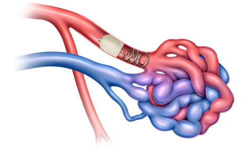
An AVM is an abnormal tangle of blood vessels where arteries connect directly to veins without the normal capillary network. This causes high-pressure blood flow that can weaken vessel walls and increase the risk of rupture and bleeding in the brain.
What is an AV Fistula?
An AV fistula is an abnormal direct connection between an artery and a vein. In the brain, these abnormal connections can occur in the dura (covering of the brain) and may cause symptoms due to increased blood pressure in veins or abnormal blood circulation.
Symptoms of AVM & AV Fistula
Symptoms may vary depending on the size and location of the vascular abnormality. Common symptoms include:
- Severe or sudden headaches
- Seizures
- Weakness or numbness in limbs
- Vision problems
- Difficulty speaking or confusion
- Brain hemorrhage in severe cases
Diagnosis
Advanced imaging techniques are used to detect and evaluate AVMs and AV fistulas.
- CT Scan – Detects brain bleeding or abnormalities
- MRI Scan – Provides detailed imaging of brain tissue and blood vessels
- CT Angiography (CTA) – Visualizes abnormal vascular structures
- Cerebral Angiography – Gold standard test to map abnormal blood vessel connections
Endovascular Embolization Procedure
During the embolization procedure, a thin catheter is inserted through a blood vessel in the groin or wrist and guided to the abnormal blood vessel using real-time imaging. Once the catheter reaches the AVM or AV fistula, special materials such as medical glue, coils, or embolic agents are injected to block the abnormal connection.
This reduces abnormal blood flow and prevents the risk of rupture or bleeding.
Benefits of Endovascular Embolization
- Minimally invasive treatment approach
- No open brain surgery required
- Reduced risk of complications
- Shorter hospital stay
- Faster recovery time
- Effective control of abnormal blood flow
Post-Treatment Care
After embolization, patients are monitored to ensure stable blood flow and proper recovery. Follow-up imaging studies may be recommended to evaluate treatment success and detect any remaining abnormal vessels.
Final Thoughts
AVM and AV fistula embolization is a highly advanced neurointervention procedure that helps treat abnormal blood vessel connections safely and effectively. With modern endovascular techniques, doctors can prevent serious complications such as brain hemorrhage and stroke while ensuring faster recovery and improved neurological outcomes.